Through continued investment, we have always been able to equip our Optometrists with the latest in eye examination and testing technologies. If we can more comprehensively gather information about your eyes, we can optimise how we assess, appraise and manage your eyes’ health and well-being. Quite simply, the more we know, the better clinicians we can be. Our Optometrists have technology on their side.
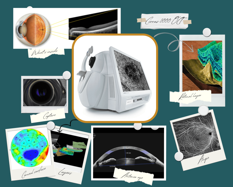
Cirrus 5000 OCT
The Zeiss Cirrus 5000 Optical Coherence Tomographer, or OCT is at the forefront of optical imaging. It allows us to view part of the eye not visible by any other means. Using a long wavelength beam of light it rapidly gathers multiple cross-sectional scans of the area in question. Each provides a slice view of the retina or optic nerve, which when blocked together create a fabulous 3D image. This image can then be virtually dissected to reveal, inspect and analyse the 13 layers that make up the inner workings of the retina. The retina itself is only the thickness of silk. This is incredible detail. Moreover, it takes a matter of seconds to complete, doesn’t involve eye drops and is pain-free.
The Cirrus 5000 OCT scans are part of our Comprehensive Eye Examination.
More Info
Using Zeiss’ Forum software we can then dovetail the information from the Cirrus with its sister device – the Clarus 500 ultra-wide field camera. We can then apply various analytical tools within the software to compare both the devices’ findings with normalative data at that time. The clever bit is then the AI guidance it can offer to compare scans time on time – to highlight changes to the retina or optic nerve that could be due to an emerging eye disease. Early detection generally = better outcomes.
Who benefits from OCT?
An OCT (Optical coherence topography) retinal scan is similar in principle to an MRI scan of the body, but uses light rather than magnetic resonance to gather its information. It provides a 3D view of the inside of the retina, not visible by any other means.
Family history of Glaucoma
It contains analytical tools that allow us to quantify what is going on with the optic nerve head – the region within the eye that is adversely affected by the Glaucoma process. Subsequent scans then, over time, highlight at the earliest stage changes that can herald the emergence of Glaucoma.
Short-sighted patients
Myopia, or short-sighted eyes, carry an increased risk of certain eye conditions, including Glaucoma, but also macular eye disease. A scan of the macular region allows a detailed inspection of microscopic internal layers of this part of the eye, allowing the Optometrist to spot potentially treatable macular conditions at the earliest of stages.
The ageing eye
All of us, simply by virtue of time, are at a higher risk of developing retinal conditions as we progress through life. At the same time, gradually the lens of our eyes loose their clarity (culminating in cataract), making the clinician’s view of the internal eye more challenging. The OCT continues to offer an accurate assessment of the central internal eye, that remains remarkably good when many other examination techniques start to fail due to this clouding up of the eye’s own lens.
All of us
The clinical insight that OCT scans provide are of relevance to us all. Maybe not now, but for the future. The more retinal/optic nerve information we can capture and record now, the longer your personal clinical data trail is, and the more compelling variances in these structures are. Eye disease, like many other conditions is about spotting change. OCT scan history allows us to do this most effectively.
OCT is included in our Advanced and Comprehensive Eye Examinations.
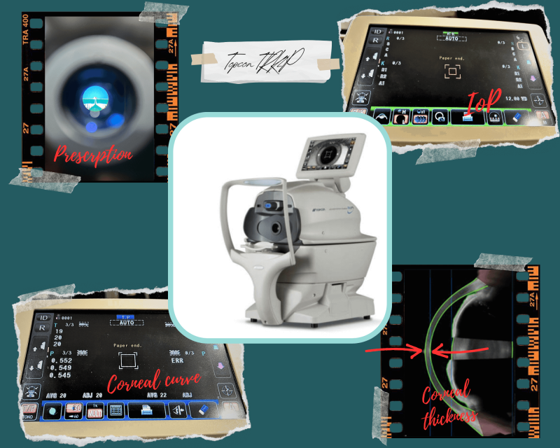
TRK 2P
The TRK 2P is a true workhorse of the examination room. It is at least four devices within one instrument and is included in our Comprehensive Eye Examination.
Kerotometer mode measures the curve of the cornea – the outer section of the eye.
Auto-Refractor mode allows us to get an objective measure of the approximate spectacle prescription.
Pachymetry mode assesses the thickness of the centre of the eye’s clear outer shell – the cornea.
Inter Ocular Pressure or IOP mode assesses the pressure of the internal eye pressure – “The Puff Test”.
Kerotometer
Most people’s eyes have a cornea that has two different curvatures – making it slightly egg-shaped – having two curves, with one curve more rounded that the other. We like to track the corneal curve for several reasons; as the eye develops through teenage years,
should the prescription unexpectedly alter, and as an ongoing part of contact lens patient care.
Auto-Refracter
It reads the de-focus of light bouncing back from an image it has just sent to the retina. The device reads how out of focus this bounce-back light is, makes adjustments and recognises the point at which focus is made, thereby determining the approximate power of the eye. Voila, the approximate prescription. You could never make glasses from this, but it can be a good starting point.
Pachymetry
As light passes from air to cornea, and then cornea to the liquid of the anterior chamber, minuscule reflections are created due to the difference in their optical density or refractive index. These are known as Purkinje Images. This device has the sensors with sufficient sensitivity to spot these two reflections, identify their separation and then use this information to deduce the corneal thickness. This is generally around 550microns, but differs between individuals.
Corneal thickness can influence the accuracy of the eye pressure ‘puff test’, so we use this measurement to more accurately calibrate the taken readings, to actual readings.
Inter Ocular Pressure
The eye has a certain level of internal pressure, which varies from individual to individual, generally increasing gradually over life. It is held in balance between the amount of liquid the eye allows into its front chamber through micro-filtration, versus the speed with which this liquid flows through the pupil to be re-absorbed back in to the body’s main circulation. This liquid is known as the aqueous. It is a highly filtered derivative of blood and its job is to bring nutrition into the eye for the cornea and lens – the only parts of the eye without their own blood supply – they need to be transparent to enable us to view through, of course!
The IOP level plays an important role in the Glaucoma process. Glaucoma is a relatively common condition to which we become more prone as we age – symptoms-free in its early stages. By measuring the IOP (along with other factors) routinely, we can screen patients for potential Glaucoma.
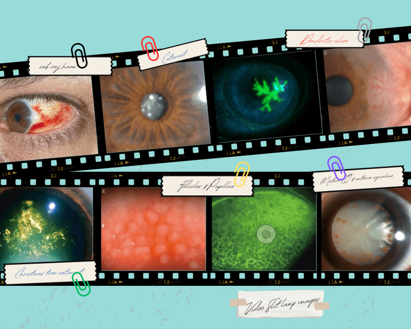
Digital LED Illuminated Slit-Lamp
This device allows an extremely clear view of the outermost structures of the eye. It has a new Infra Red illumination feature allowing an assessment of one of the glands of the eye that produces tear oils – otherwise invisible through standard systems. The digital slit lamp also allows us to show you views of your eye on the remote TV screen and record feature for later comparison if necessary.
This is included in our Comprehensive Eye Examination.
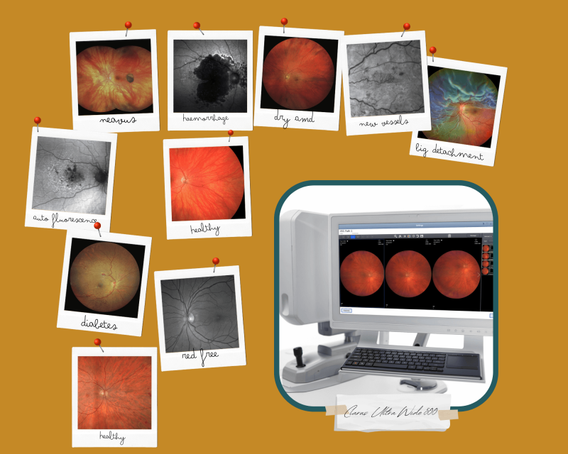
Clarus 500
Zeiss’ Clarus 500 is the next generation of ultra-widefield retinal imaging. Until quite recently, if you wished to capture an image of anything on the retina beyond a 45 degree spread, you would have to use pupil enlargement drops to achieve this. This variety of eyedrop allows a fantastic view in the eye, but isn’t ideal for routine checks due to unwanted, lingering effects of dilation – namely blurred vision and increased sensitivity to light.
The more of the eye we can see, the better guided clinical assessments and decisions can be made. The Clarus enables this.
Widefield retinal imaging is included in our Comprehensive Eye Examination.
More Info
The Clarus 500 camera in a single take, without any eye drops, provides an image 130 degrees wide. This increases to an ultra widefield 200 degrees with two takes, using some nifty software to stitch the two images together. A further ‘montage’ mode pushes this out to over 260 degrees. It literally has the ability to see around corners.
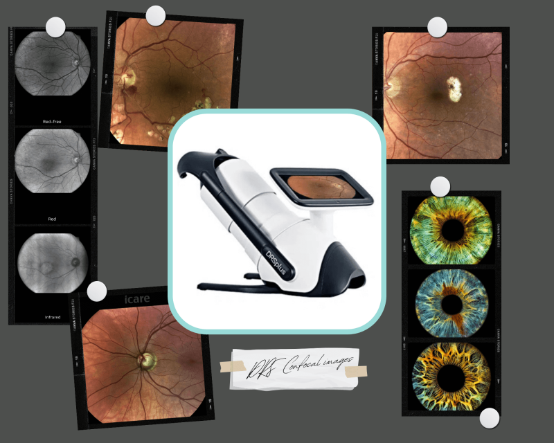
DRS
The I-Care DRS Plus is a retinal imaging system and is quite a device. It uses either one or two captures, with some clever photo montaging software, to provide a high quality, high resolution, detail-rich image of the retina.
You will be surprised to see how much is in there – there is a lot to see, and it can give us insights into many other aspects of your general health.
The DRS plus is used as part of our Standard Private Examination.
More Info
The examination is comfortable – the wavelength of light used enters and leaves the eye with the softest of flashes. These images are then beamed to the examination room. Our Optometrist will then take time to guide you through what we see in your retinal image, along with how and where elements can change with common eye conditions. If you’ve had previous images at your last test, there is some impressive software that can overlay current and previous images, thereby assisting us in spotting these subtle changes.
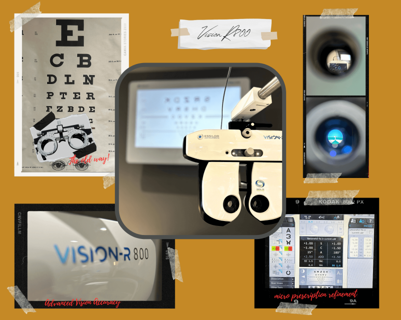
Vision R800
The Vision R800 is the newest reinvention of the most iconic part of the eye examination – the part that works out how easily your eyes focus and align, providing your “prescription” or refractive error measurement. Gone are the uncomfortable optician glasses and hand lenses, and instead, this….
A digital refraction process allows us to get the best out of your eye’s focusing system, with an efficient, precise procedure.
It is comfortable, as it floats above your nose, and your vision is assessed with changes in lens strength almost magically in front of your eyes, as you gaze through towards the letter chart.
This is included in our Comprehensive Eye Examination.
More Info
Traditionally, an eyesight test would involve using lenses that step up and down in 0.25D increments. Research by the R800’s developers found that 95% people have more sensitive vision than the current 0.25 correction steps – steps per diopter of power. The optical module they developed allows adjustment in power to 0.01D – 100 steps per diopter. The resulting prescription then allows us to most thoroughly unlock your eye’s seeing potential. This is the Advanced Vision Accuracy (AVA) prescription, from which bespoke lenses can be manufactured integrating this precise prescription. A true Wow!
The Vision R800 is part of the Comprehensive Eye Examination.
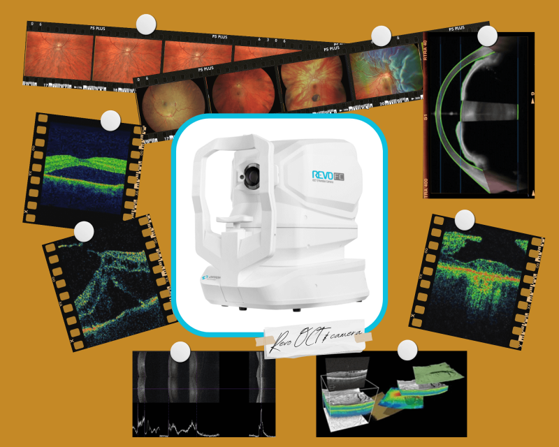
Revo
The Revo FC is an incredibly powerful and versatile new device in our armoury. Developed by famed technology innovators Optopol, it saddles the combinations of a 45degree 12.3 mega pixel camera, with a Spectral domain OCT, Glaucoma & retinal analysis, Biometer and Corneal topographer in one device.
This is the perfect device for checking and monitoring the eye’s structure and function over time – particularly useful in the identification of the earliest indicators of Glaucoma and Macular disorders.
This is included in our Advanced Eye Examination.
Who benefits from retinal photography?
Everyone! A photograph of the internal eye provides a permanent reference point in time. A record of the retinal pigmentation and complexion, the way the tiny blood vessels fan out and interact over the retinal surface and the features of the visible part of the optic nerve. All of these elements are unique to you. A photograph can then be compared for future reference.
Retinal photography is included in our Private Essential Eye Examination.
Who benefits from OCT?
An OCT (Optical coherence tomography) retinal scan is similar in principle to an MRI scan of the body, but uses light rather than magnetic resonance to gather its information. It provides a 3D view of the inside of the retina, not visible by any other means.
Family history of Glaucoma
It contains analytical tools that allow us to quantify what is going on with the optic nerve head – the region within the eye that is adversely affected by the Glaucoma process. Subsequent scans then, over time, highlight at the earliest stage changes that can herald the emergence of Glaucoma.
Short-sighted patients
Myopia, or short sighted eyes, carry an increased risk of certain eye conditions, including Glaucoma, but also macular eye disease. A scan of the macular region allows a detailed inspection of microscopic internal layers of this part of the eye, allowing the Optometrist to spot potentially treatable macular conditions at the earliest of stages.
The ageing eye
All of us, simply by virtue of time, are at a higher risk of developing retinal conditions as we progress through life. At the same time, gradually the lens of our eyes are loosing their clarity (culminating in cataract), making the clinician’s view of the internal eye more challenging. The OCT continues to offer an accurate assessment of the central internal eye, that remains remarkably good when many other examination techniques start to fail due to this clouding up of the eye’s own lens.
All of us
The clinical insight that OCT scans provide are of relevance to us all. Maybe not now, but for the future. The more retinal/optic nerve information we can capture and record now, the longer your personal clinical data trail is, and the more compelling variances in these structures are. Eye disease, like many other conditions, is about spotting change. OCT scan history allows us to do this most effectively.
OCT is included in our Advanced and Comprehensive Eye Examinations.
Who benefits from Biometry?
This module forms the mainstay of our Myopia Management Programme (MMP). It allows us to painlessly and accurately measure the length of the internal eye from front to back. This is referred to as the eye’s Axial Length. We use this to check on how effectively we are keeping a child’s myopia under control. There is value however to knowing all children’s axial length, as there are increased risks of eye disease in certain eye ‘sizes’, which can be potentially mitigated by structured on-going eyecare for life.
Biometry is included in MMP fees as standard, but available as an additional test for NHS funded under 16s.
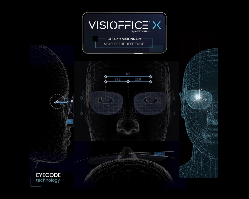
VisiOffice X
Once you have had your examination, your optical needs discussed and stylish and suitable frames selected, the next step is measurement. Traditionally, this is where the trusted small frame ruler would be used to measure various aspects of frame fit and eye position in that frame. Things have moved on. Meet the VisiOffice X. An exceptional optical measuring system. Access to truly bespoke lenses.
Find out More
- Armed with three cameras working simultaneously, the VisiOffice gives us a three dimensional reconstruction of the eye, frame and lens combination
- 11 different eye/frame/face parameters are assessed
- It looks at the individual pupil spacing both vertically and horizontally using the reflection off the corneal surface. You’ll be surprised how much asymmetry there is in our faces
- The frame’s distance from the front of the eye is measured, along with its curve relative to your face
- It will have a specific tilt angle according to how high or low your ears are positioned, this is also measured
So far, maybe familiar. Next we have the clever bit.
By watching how you look in different directions, it then makes an assessment of the eye’s centres of rotation – how deep the orbits are, and where the eye is in the optical equation. Finally, we have a near vision assessment. VisiOffice watches you perform a near vision task, watching and learning about your Near Vision Behaviour. How much do you drop your head? How much do you turn your face? How much eye movement versus head movement do you make? Do you converge your eyes evenly? Do you have a dominant eye? All measured.
These elements and factors are then number-crunched to create your personal Eye Code, which allows us to personalise your spectacle lens. Unique to not only your spectacle prescription, your eyes’ centre of rotation, but also the chosen frame and how it sits on your face.
The spectacle lens is then designed for you. Bespoke and personal.
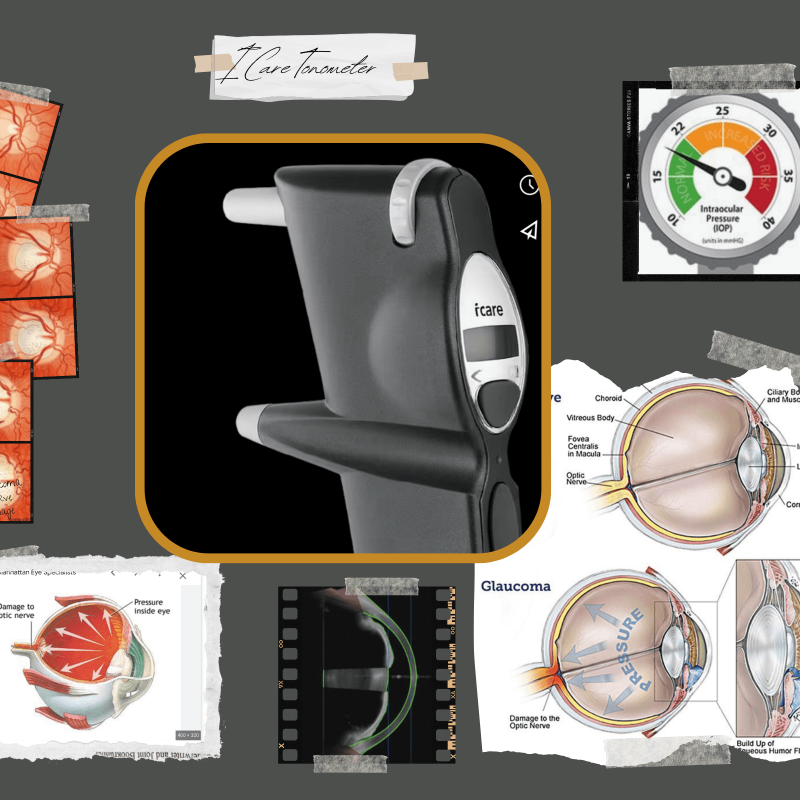
iCare Tonometry
For those that dislike the traditional means of measuring eye pressure we offer an alternative. This device uses a state of the art magnetic, floating probe which flickers against the tear film with barely a sensation. This can be used in either our Standard or Comprehensive Examinations.